Conditions treated
Interventional radiology may be used to manage vascular blockages, abnormal bleeding, blood clots, varicose veins, dialysis-access problems, selected tumours and abnormal fluid collections.
Fast, precise imaging and image-guided treatment. Our team combines clinical depth, coordinated diagnostics and clear communication so every patient understands their options.
Radiology plays an important role in detecting diseases, confirming diagnoses, planning treatment and monitoring recovery. Interventional radiology goes a step further by using medical imaging to guide minimally invasive procedures inside the body.
The Department of Radiology & Interventional Radiology at ANU Hospitals, Vijayawada, supports patients and referring doctors with diagnostic imaging and image-guided interventions. Our focus is on accurate evaluation, appropriate procedure selection, patient safety and coordinated care across multiple medical specialities.
Patients searching for a radiologist in Vijayawada can consult the department for imaging evaluation, image-guided procedures and specialist interpretation. Those searching online for the best interventional radiologist in Vijayawada should consider the doctor’s qualifications, experience, relevant procedural expertise, hospital support and whether the recommended treatment is appropriate for their condition—not promotional claims alone.
Radiology is a medical speciality that uses imaging technologies to examine structures and organs inside the body. Radiologists are qualified medical doctors trained to interpret images, identify abnormalities and communicate findings to the treating doctor.
Different imaging methods provide different types of information. An X-ray may help evaluate bones and lungs, ultrasound uses sound waves to examine soft tissues and blood flow, and CT and MRI can provide detailed cross-sectional images of internal organs.
The radiologist selects or recommends the most appropriate imaging method according to the patient’s symptoms, medical history and the clinical question that needs to be answered. Not every patient requires every type of scan.
Interventional radiology is a specialised branch of radiology in which doctors use real-time imaging to guide small instruments, needles, wires or catheters through the body. These procedures are commonly performed through a small skin puncture instead of a large surgical incision.
Ultrasound, fluoroscopy, CT or other imaging methods may be used to guide the procedure accurately. Depending on the condition, interventional radiology may help open a narrowed blood vessel, stop internal bleeding, obtain a tissue sample, drain an abnormal fluid collection or block the blood supply to a selected area.
Many image-guided interventions can be performed under local anaesthesia with sedation when appropriate. However, suitability, potential benefits, risks, alternatives and expected recovery must be assessed individually by the interventional radiologist and referring specialist.
The department works with specialists in neurology, cardiology, gastroenterology, nephrology, urology, oncology, orthopaedics, vascular care, general surgery, gynaecology, critical care and emergency medicine.
Radiology services may assist with:
The investigation or procedure recommended for a patient depends on their symptoms, diagnosis, previous reports, kidney function, allergies, medications and general health.
Interventional radiology may be used to manage vascular blockages, abnormal bleeding, blood clots, varicose veins, dialysis-access problems, selected tumours and abnormal fluid collections.
It can also support the treatment of uterine fibroids, enlarged prostate, vascular malformations and conditions requiring biopsy, drainage or image-guided catheter placement.
Not every condition is suitable for an intervention; eligibility is determined after clinical examination and review of relevant scans and test results.
The availability and suitability of a particular procedure must be confirmed directly with the department.
Symptoms vary according to the organ or blood vessel involved and may include limb pain, swelling, colour changes, non-healing wounds, unusual bleeding or persistent abdominal discomfort.
Women with heavy menstrual bleeding or pelvic pressure and patients with sudden limb swelling or reduced circulation may require specialist assessment.
These symptoms can have many causes and do not confirm a specific disease; urgent or persistent symptoms should be evaluated by a qualified doctor.
Other symptoms that may lead to radiology or interventional radiology evaluation include:
Sudden severe pain, uncontrolled bleeding, weakness on one side of the body, difficulty speaking, chest pain, breathlessness or a cold and painful limb may represent an emergency. Seek immediate medical assistance rather than waiting for an outpatient appointment.
Diagnosis begins with a review of symptoms, medical history, previous scans, laboratory results and the referring doctor’s clinical findings.
The radiologist may use X-ray, ultrasound, Doppler, CT, MRI or other appropriate imaging to identify an abnormality and understand its location and extent.
When imaging alone cannot confirm the diagnosis, an image-guided biopsy, aspiration or another targeted procedure may be recommended.
X-rays are commonly used to examine bones, joints and the chest. They may help identify fractures, infections, lung abnormalities and certain degenerative changes.
Ultrasound uses sound waves rather than ionising radiation. It may be used to examine the abdomen, pelvis, thyroid, breast, soft tissues and selected organs.
Doppler ultrasound evaluates blood flow through arteries and veins. It may help detect narrowing, blockage, venous reflux or blood clots.
CT creates detailed cross-sectional images of the body and may be used in trauma, emergency assessment, cancer evaluation, vascular studies and the examination of internal organs. Contrast material may be needed for some scans.
MRI uses magnetic fields and radio waves to create detailed images. It can be useful for evaluating the brain, spine, joints, soft tissues and selected internal organs.
Angiography provides detailed images of blood vessels. It may be performed for diagnosis, treatment planning or as part of an interventional procedure.
A radiologist may use ultrasound or CT guidance to place a needle accurately into an abnormal area and collect tissue or fluid. The sample is then examined by a pathologist.
The radiologist interprets imaging findings in the context of the patient’s clinical information. Imaging findings should be discussed with the treating doctor because a scan report alone may not provide the complete diagnosis.
Treatment may involve angioplasty, stenting, embolisation, thrombus management, drainage, biopsy or another image-guided minimally invasive procedure.
A thin catheter or needle is directed to the treatment area using imaging, often through a small puncture in the skin.
The recommended procedure depends on the diagnosis, imaging findings, expected benefits, available alternatives and the patient’s overall condition.
Angioplasty may be used to open a narrowed or blocked blood vessel with a small balloon. A stent may be placed in selected cases to help keep the vessel open.
These procedures may be considered for certain patients with peripheral arterial disease or other vascular narrowing. Suitability depends on the location and severity of the blockage, symptoms and overall health.
Embolisation involves intentionally blocking blood flow to a selected blood vessel or treatment area. It may be used to control bleeding, manage certain vascular abnormalities or reduce the blood supply to selected tumours or uterine fibroids.
The material used and the extent of embolisation depend on the condition. The doctor explains the treatment objective and potential risks before obtaining consent.
Uterine artery embolisation may be considered for selected women with symptomatic uterine fibroids. The procedure blocks selected blood vessels supplying the fibroids, which may help reduce their size and associated symptoms.
A gynaecological assessment and appropriate imaging are usually required before determining suitability. Fertility plans and alternative treatments should be discussed with the relevant specialists.
Some patients with extensive or severe deep vein thrombosis may be evaluated for catheter-directed treatment. Options can vary according to the location and duration of the clot, bleeding risk and severity of symptoms.
Most blood clots are treated medically. An interventional procedure is recommended only when the treating team believes it may provide an appropriate benefit.
Image-guided treatment may be considered for selected patients with painful varicose veins, swelling, skin changes or venous ulcers. Doppler ultrasound is commonly used to understand the pattern of venous reflux before treatment planning.
The appropriate option depends on the affected veins, symptoms, previous treatments and individual risk factors.
Patients undergoing haemodialysis may develop narrowing, clotting or reduced flow within an arteriovenous fistula or graft. Imaging and catheter-based procedures may help identify and treat selected access problems.
Early evaluation may help preserve dialysis access, but the urgency and treatment approach must be decided by the dialysis and vascular-care teams.
Ultrasound or CT may be used to guide a small tube into an abscess or abnormal collection. This can allow infected or unwanted fluid to drain while reducing injury to nearby structures.
Antibiotics, surgery or additional treatment may still be required depending on the underlying condition and response.
Image-guided procedures may help relieve selected blockages affecting the bile ducts or urinary system. A drain or stent may be placed when clinically appropriate.
These procedures are planned in coordination with gastroenterologists, surgeons, urologists or other relevant specialists.
Radiology uses technologies such as digital X-ray, ultrasound, Doppler, CT, MRI, fluoroscopy and angiographic imaging to examine internal structures and guide care.
Interventional radiology uses real-time imaging with specialised needles, wires, catheters, balloons, stents and embolisation materials for selected procedures.
The technology used for an individual patient is chosen according to the clinical need, safety considerations and the type of intervention planned.
Technology is only one part of safe radiology care. Accurate interpretation, appropriate patient selection, radiation safety, sterile technique, careful monitoring and communication between specialists are equally important.
The American College of Radiology defines radiologists as medical doctors who diagnose and treat disease using imaging examinations and image-guided procedures.
Depending on the procedure and the patient’s condition, potential benefits may include:
These are potential benefits, not guarantees. Some patients may still require surgery or another treatment. Recovery and outcomes vary according to the condition, procedure and individual risk factors.
Before an interventional procedure, the doctor may review blood tests, kidney function, clotting results, allergies and current medications. Patients should inform the team if they:
Do not stop prescribed medicines unless the treating doctor instructs you to do so. Fasting, admission and medication instructions vary by procedure.
Radiological procedures are performed only after considering their expected clinical benefit. When ionising radiation or contrast material is needed, the team takes appropriate precautions and follows relevant safety protocols.
Recovery instructions depend on the procedure performed. Some patients may return home after observation, while others require hospital admission and monitoring.
Patients may be advised to:
Seek immediate medical attention for uncontrolled bleeding, breathing difficulty, severe pain, loss of consciousness, new weakness, a cold or discoloured limb or another rapidly worsening symptom.
ANU Hospitals provides diagnostic and interventional radiology support within a multispeciality hospital environment. This enables the radiology team to coordinate with physicians, surgeons, emergency doctors and critical-care specialists when a patient requires combined evaluation and treatment.
The department includes Dr. Bhavani Shankar Manam, Consultant Endovascular & Interventional Radiologist, with qualifications listed as MBBS, MD, DNB (Radiodiagnosis) and FIVR. His profile, medical registration information and areas of expertise should be displayed clearly on the website to strengthen patient trust and E-E-A-T signals.
When choosing a radiologist in Vijayawada, patients should look for verified qualifications, relevant experience, transparent explanations, hospital support and a treatment plan based on clinical need.
A diagnostic radiologist interprets medical images to help identify diseases and injuries. An interventional radiologist also performs minimally invasive procedures using imaging to guide needles, wires and catheters inside the body.
No. It may offer an alternative for selected conditions, but it cannot replace surgery in every case. The most appropriate treatment depends on the diagnosis, disease severity and the patient’s overall health.
Many procedures are performed under local anaesthesia, sometimes with sedation. Patients may feel pressure or temporary discomfort, but pain-control methods depend on the procedure and individual requirements.
Some procedures can be completed with short observation, while others require admission. The doctor will explain the expected hospital stay during pre-procedure counselling.
A referral is helpful because it provides the clinical reason for imaging or intervention. Patients seeking a second opinion should bring their prescriptions, scan images, reports and previous treatment records.
Avoid relying only on promotional rankings. Check the doctor’s recognised qualifications, medical registration, relevant procedural experience, hospital facilities, approach to patient safety and willingness to explain benefits, risks and alternatives.
If you need diagnostic imaging, an image-guided biopsy, vascular evaluation or an opinion about a minimally invasive procedure, consult the Radiology & Interventional Radiology team at ANU Hospitals.
Appointment number: 070706 49999
Interventional radiology may be used to manage vascular blockages, abnormal bleeding, blood clots, varicose veins, dialysis-access problems, selected tumours and abnormal fluid collections.
ymptoms vary according to the organ or blood vessel involved and may include limb pain, swelling, colour changes, non-healing wounds, unusual bleeding or persistent abdominal discomfort.
Diagnosis begins with a review of symptoms, medical history, previous scans, laboratory results and the referring doctor’s clinical findings.
Treatment may involve angioplasty, stenting, embolisation, thrombus management, drainage, biopsy or another image-guided minimally invasive procedure.
Radiology uses technologies such as digital X-ray, ultrasound, Doppler, CT, MRI, fluoroscopy and angiographic imaging to examine internal structures and guide care.
Structured follow-up, rehabilitation support and preventive guidance for sustainable recovery.

Radiology & Interventional Radiology
View profile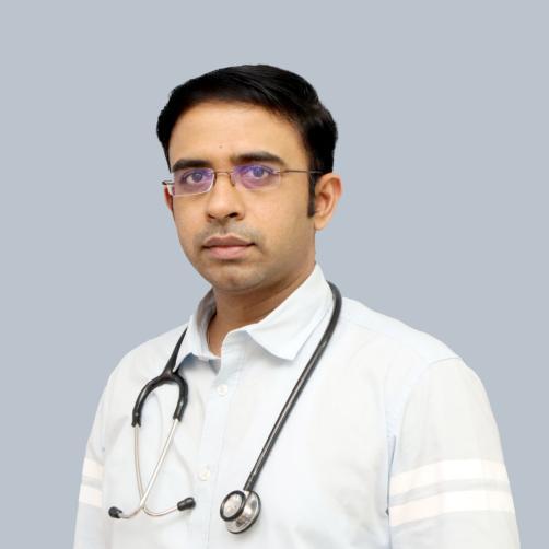
Radiology & Interventional Radiology
View profileAppointments, emergencies and second opinions???our care team is here 24/7.